Tuesday, April 6, 2021

Newly discovered antibiotics kill bacteria differently

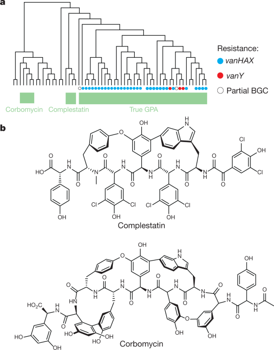
Image result for corbomycin structure


Antibiotic resistance is a growing public health problem across the globe, with many diseases becoming harder to treat. Now, a newly discovered antibiotic group shows promise in the fight against superbugs as it has a unique way of killing bacteria.
A team of scientists at McMaster University has found a new group of antibiotics that can fight infections in a new and unique way. These antibiotics fight infections in a way researchers have never seen before, according to the findings of the study described in the journal Nature.

'Holy grail' of antibiotics

The newly found group of antibiotics, consisting of corbomycin and complestatin, can kill bacteria by blocking the function of the bacterial cell wall. These drugs come from a family of antibiotics known as glycopeptides, which are produced by soil bacteria.
The two antibiotics attack peptidoglycan, the main component of the bacterial cell wall that is vital to the growth and survival of almost all bacteria. They inhibit the action of autolysins, which are important for cell division and growth.
Other antibiotics, such as penicillin, work by preventing the bacteria from building its wall, which is the source of its strength. In killing the bacteria, removing its wall will make it vulnerable and easier to kill.
These new antibiotics work by doing the opposite. Instead of preventing building the wall, it halts the wall ll from being broken down. As a result, blocking the breakdown of the wall would make it impossible for them to divide and expand – just like being trapped in prison.

Unique bacteria killer

The two new antibiotics are known as glycopeptides. The team studied the genes of the group to see if they lack resistance mechanisms. The team believes that if the genes that made these drugs different, perhaps the way they kill will also be different.
In collaboration with scientists from the Université de Montréal, including Yves Brun, they found that the drugs act on the bacterial cell wall to prevent it from dividing and proliferating.  
"Knowing the detailed structure at the atomic level of this connection between the surface layer and the surface of the cell offers enormous potential to then develop molecules that can target this attachment and make the cell more sensitive to antibacterials," Yves Brun, study co-author, said.
"Combined with the discovery of the new mode of action of two antibiotics, this development opens up prospects for weakening the action of bacteria and making them more vulnerable," he added.
The researchers believe the group of drugs is a promising clinical candidate in the hopes of stemming bacteria from becoming resistant to antibiotics.

Fight against antibiotic resistance

Antibiotic resistance is one of the greatest threats to global health, according to the World Health Organization (WHO). Though it happens naturally, the misuse of antibiotics is hastening the process, making it easy to treat infections in the past harder to curb now.
Further, antibiotic resistance increase hospital stays and medical costs. For instance, diseases in the past that were responsive to certain antibiotics may become resistant and difficult to stem, such as tuberculosis, pneumonia, gonorrhea, and other infections. Now, as the diseases become stronger and more resilient, outbreaks may become inevitable, unless new drugs are discovered.
In the United States alone, at least 2.8 million people become infected with antibiotic-resistant bacteria each year, while more than 35,000 people die.

https://www.nature.com/articles/s41586-020-1990-9

Monday, April 5, 2021

Novel drug combination discovered to induce high rates of human beta cell proliferation


Harmine structure.svg

Harmine

Researchers at the Icahn School of Medicine at Mount Sinai have discovered a novel combination of two classes of drugs that, together, cause the highest rate of proliferation ever observed in adult human beta cells- the cells in the pancreas that produce insulin- without harming most other cells in the body. The result is an important step toward a diabetes treatment that restores the body's ability to produce insulin.
The finding involved one type of drug that is known to cause beta cells to proliferate and another that is already in widespread use in people with diabetes. Together, they caused the cells to proliferate at a rate of 5 to 6 percent per day. The study was published today in Science Translational Medicine online.

We are very excited about this new drug combination because for the first time ever, we are able to see rates of human beta cell replication that are sufficient to replenish beta cell mass in humans with diabetes."
Andrew Stewart, MD, Director of the Mount Sinai Diabetes, Obesity, and Metabolism Institute and lead author of the study
Diabetes occurs when there are not enough beta cells in the pancreas, or when those beta cells secrete too little insulin, the hormone required to keep blood sugar levels in the normal range. Approximately 30 million people in the United States have diabetes and nearly 50 to 80 million more are living with prediabetes (also called "metabolic syndrome"). Diabetes can lead to major medical complications: heart attack, stroke, kidney failure, blindness, and limb amputation.
In type 1 diabetes, the immune system mistakenly attacks and destroys beta cells. A deficiency of functioning beta cells is also an important contributor to type 2 diabetes, the most common type of diabetes. Thus, developing drugs that can increase the number of healthy beta cells is a major priority in diabetes research.
According to Dr. Stewart, none of the diabetes drugs currently on the market can induce beta cell regeneration in people with diabetes. In parallel with the Mount Sinai work, other researchers are studying pancreatic transplantation, beta cell transplantation, and stem cell replacement of beta cells for people with diabetes, but none of these approaches is in widespread use.
"This is a very exciting discovery in the field of diabetes and is a key next step in drug development for this disease," said Dennis S. Charney, MD, Anne and Joel Ehrenkranz Dean, Icahn School of Medicine at Mount Sinai. "This important work truly holds promise for so many people."
In 2015, Dr. Stewart and his team published a paper in Nature Medicine that showed that harmine, a drug that inhibits the enzyme dual specificity tyrosine-regulated kinase 1A (DYRK1A), induced multiplication of adult human beta cells. In that study, his team also discovered that harmine treatment led to normal control of blood sugar and proliferation in human beta cells in diabetic mice whose beta cells had been replaced with small numbers of transplanted human beta cells. While this was a major advance, the proliferation rate was lower than needed to rapidly expand beta cells in people with diabetes.
This current paper builds upon a study that Dr. Stewart and his team published in Cell Metabolism in December 2018 where they discovered that DYRK1A inhibitors combined with another drug that inhibits transforming growth factor beta superfamily members (TGFβSF), also known as a family of proteins with various biological processes such as growth, development, tissue homeostasis and immune system, could cause beta cells to proliferate at a rate of 5 to 8 percent per day. However, according to Dr. Stewart, TGFβSF's would likely have side effects on other organs in the body that would prevent clinical use.
The next challenge was developing ways to target regenerative drugs to the beta cells while avoiding other cells and organs in the body where they may elicit adverse effects.
In the study published today, titled "GLP-1 receptor agonists synergize with DYRK1A inhibitors to potentiate functional human beta cell regeneration," Dr. Stewart and his team combined DYRK1A inhibitors like harmine with a class of beta cell-targeting drugs, also known as GLP1R agonists, which are already in widespread use in people with type 2 diabetes. They showed-;in beta cells from normal people and people with type 2 diabetes, both in the tissue culture dishes and in human beta cells transplanted into mice-;that combining harmine (or any other DYRK1A inhibitor) with any of the many GLP1R agonist drugs currently on the market for diabetes yields high rates of human beta cell replication, and does so in a way that is highly selective for the beta cell.
The project arose from the PhD thesis of an Icahn School of Medicine graduate student, Courtney Ackeifi, now a postdoctoral fellow in Dr. Stewart's lab and first author of the paper, who explored a broad spectrum of potential drug partners that could enhance the beta cell regenerative efficacy and selectivity of harmine.
Said Dr. Ackeifi of the discovery, "The beauty here is that the combination of DYRK1A inhibitors with GLP1R agonists achieves the highest rate of human beta cell replication possible, and does so in a highly specific way. This is an important advance in the field of diabetes because we may have found a way to convert a widely used class of diabetes drugs into a potent human beta cell regenerative treatment for all forms of diabetes."
"We know that a critical pathway to drive a cure for type 1 diabetes includes transplanting insulin-producing beta cells into people or enticing their existing beta cells to start multiplying," explains Francis Martin, PhD, JDRF Director of Research. "It is exciting to learn from the work of Dr. Stewart and his team that GLP1R agonists could increase the effect of the recently discovered agents that promote multiplication. Using GLP1R offers a means to boost the effect while also improving the safety of this type of drug."
The next goals of the project are to perform long-term studies in animals transplanted with human beta cells, and to determine if any cells or organs in the body other than beta cells are affected by the new drug combination.
https://en.wikipedia.org/wiki/Harmine
https://en.wikipedia.org/wiki/Glucagon-like_peptide-1_receptor_agonist

Friday, April 2, 2021

Oligosaccharide 2'FL in breast milk enhances cognitive development in babies

2'-fucosyllactose Drawing.png


Maternal factors, such as breast milk, have been shown to affect a baby's development, and previous animal studies have determined that a carbohydrate, the oligosaccharide 2'FL found in maternal milk, positively influences neurodevelopment. Now, in the first study done in humans, investigators at Children's Hospital Los Angeles in collaboration with the University of California, San Diego, have shown that 2'FL found in breast milk enhances cognitive development. Findings will be published in PLOS ONE on Feb 12.
In this cohort study of 50 mothers and their babies, investigators analyzed breast milk composition and frequency of feeding at 1 and 6 months of age. Cognitive development was measured at 24 months using the Bayley-III scale, a standardized test of infant and toddler development. The study showed that the amount of 2'FL in breast milk in the first month of feeding was related to significantly higher cognitive development scores in babies by 2 years of age. The amount of 2'FL in breast milk at 6 months of feeding was not related to cognitive outcomes, indicating that early exposure may be more beneficial.
Many studies have reported a positive effect of breastfeeding on cognitive development. "We wanted to specifically identify what was causing this effect," said Michael Goran, PhD, Director of the Diabetes and Obesity Program at The Saban Research Institute of Children's Hospital Los Angeles and senior author on the study.
Through our high-throughput analytical platform we can quantify oligosaccharides like 2'FL and many others in hundreds of breast milk samples in a short period of time. This technology allows us to associate differences in milk composition with specific infant outcomes like cognitive development, validating existing data from preclinical models or generating entirely new hypotheses."
Lars Bode, PhD, study collaborator and co-author, Professor of Pediatrics and Director of the Mother-Milk-Infant Center of Research Excellence at the University of California, San Diego

Using a statistical technique called mediation analysis, the investigators were able to independently evaluate the effects of breastfeeding in general, and the effects of the oligosaccharide 2'FL.
"This enhanced cognitive development in the first 2 years of life raises the question of possible long term impact on a child¬-in school and beyond," said Paige Berger, PhD, RD, a postdoctoral research associate at CHLA and the first author of the study.
These observations allowed the team to conclude that the increased neurodevelopment provided by breastfeeding was due primarily to mothers who were producing more 2'FL for the baby to consume.
"We know that there are many different compounds in breast milk and the composition is dynamic - it changes over time and is highly variable between mothers," Dr. Goran said. "In addition to identifying the impact of oligosaccharide 2'FL, we also wanted to determine the timing of when it is most critical to a child's development."
While the investigators observed neuroenhancement explained by higher 2'FL during the first month of a baby's life, this effect was not observed when looking at 2'FL content of breast milk at the six-month time-point. Being able to identify factors critical to early neurodevelopment offers the possibility for supplementing women's breast milk in individuals who produce lower quantities of this important substance.
"For some women, breastfeeding is a challenge. For those that are not able to breastfeed or can only do so short-term, 2'FL could potentially be offered as an add-on to the nutrition their baby is receiving to better support cognitive development," said Dr. Berger.
https://journals.plos.org/plosone/article?id=10.1371/journal.pone.0228323
https://en.wikipedia.org/wiki/2'-Fucosyllactose

Thursday, April 1, 2021

Common drug could mitigate risk of 'a broken heart' during bereavement

In continuation of my update on aspirin
The increased risk of heart attack or "a broken heart" in early bereavement could be reduced by using common medication in a novel way, according to a world-first study led by the University of Sydney and funded by Heart Research Australia.
Lead Investigator Professor Geoffrey Tofler said while most people gradually adjust to the loss of a loved one, there is an increase in heart attack and death among bereaved people, particularly those grieving a spouse or child.
"The increased risk of heart attack can last up to six months. It is highest in the first days following bereavement and remains at four times the risk between seven days to one month after the loss."
The study, published in the American Heart Journal, is the first randomized controlled clinical trial to show it is possible to reduce several cardiac risk factors during this time, without adversely affecting the grieving process.
"Bereavement following the death of a loved one is one of the most stressful experiences to which almost every human is exposed," said Professor Tofler, Professor of Preventative Cardiology at the University of Sydney's Faculty of Medicine and Health, and Senior Staff Cardiologist at Royal North Shore Hospital.
"Our study is the first clinical trial to examine how the cardiac risk factors could be mitigated during early bereavement."

About the study

The research team from the University of Sydney, Royal North Shore Hospital and the Kolling Institute enrolled 85 spouses or parents in the study within two weeks of losing their family member.
Forty-two participants received low daily doses of a beta blocker and aspirin for six weeks, while 43 were given placebos. Heart rate and blood pressure were carefully monitored, and blood tests assessed blood clotting changes.
The main finding was that the active medication, used in a low dose once a day, successfully reduced spikes in blood pressure and heart rate, as well as demonstrating some positive change in blood clotting tendency."
Professor Geoffrey Tofler, lead investigator
The investigators also carefully monitored the grief reaction of participants.
"We were reassured that the medication had no adverse effect on the psychological responses, and indeed lessened symptoms of anxiety and depression," said Professor Tofler.
"Encouragingly, and to our surprise, reduced levels of anxiety and blood pressure persisted even after stopping the six weeks of daily beta blocker and aspirin."
Aspirin-skeletal.svg
Co-investigator Associate Professor Tom Buckley said the study builds on the team's novel work in this area with their earlier studies among the first to identify the physiological correlates of bereavement.
"While beta blockers and aspirin have been commonly used long term to reduce cardiovascular risk, they have not previously been used in this way as a short-term preventative therapy during bereavement," said
Associate Professor Buckley of the University of Sydney Susan Wakil School of Nursing and Midwifery.

Implications and next steps

The authors acknowledge that larger long-term studies are needed to identify who would benefit most however the findings provide encouragement for health care professionals to consider this preventative strategy among individuals that they consider to be at high risk associated with early bereavement.
"Our finding on the potentially protective benefit of this treatment is also a good reminder for clinicians to consider the well-being of the bereaved," said Associate Professor Buckley.
"Future studies are needed to assess if these medications could be used for other short periods of severe emotional stress such as after natural disasters or mass bereavement where currently there are no guidelines to inform clinicians."
Co-investigator Dr. Holly Prigerson, Co-Director of the Center for Research on End-of-Life Care at Weill Cornell Medicine in New York, said:
This is an important study because it shows ways to improve the physical and mental health of at-risk bereaved people. It is a preventive intervention that is potentially practice-changing, using inexpensive, commonly available medicines."
People experiencing cardiac symptoms should discuss their condition with a health care professional before taking medication as incorrect use could be harmful.
More about beta-blockers
https://en.wikipedia.org/wiki/Aspirin
https://www.sciencedirect.com/science/article/abs/pii/S0002870319303047?via%3Dihub

Wednesday, March 31, 2021

Statin use alone or with metformin may increase survival in high-risk prostate cancer patients

In continuation of my update on metformin


Image result for metformin


Among high-risk prostate cancer patients - those with high PSA and Gleason scores of 8 or more - many will develop a difficult-to-treat disease. Preliminary research suggests that two commonly prescribed medications, cholesterol-lowering statins and the diabetes therapy metformin may have anticancer effects. However, it is unclear which of these two medications - commonly prescribed together -- contributes the most and whether they can impact high-risk prostate cancer. New research shows that statins, alone or with metformin, increase survival in men with high-risk prostate cancer.
"Both metformin and statins have been associated with longer life in prostate cancer patients, yet because they are commonly prescribed together, no study we know of has looked at these two medications separately," says senior author Grace Lu-Yao, PhD, associate director of Population Science at the Sidney Kimmel Cancer Center--Jefferson Health, one of only eight NCI-designated cancer centers nationwide with a prostate cancer program of excellence.
The study, published in Cancer Medicine on Feb 8th, looked at a number of statin therapies, and metformin, an anti-diabetic medication, in high-risk prostate cancer populations.
Using data from the Surveillance, Epidemiology and End Results (SEER-18) database linked with Medicare files, Dr. Lu-Yao and colleagues looked at patients diagnosed with cancer from 2007 through to 2011. Based on 12,700 patients, the researchers observed that statins alone or in combination with metformin was significantly associated with reduced mortality from all causes.
Dr. Lu-Yao and colleagues saw the highest median survival of 3.9 months in men who took both metformin and statins, 3.6 with statins alone and 3.1 years with metformin alone. The median survival for those who did not use either drug was also 3.1 years.
With respect to prostate mortality, metformin plus statin was associated with a 36% reduction in risk of death followed by statins alone. Those taking metformin alone were relatively rare, and there was no significant association with all-cause mortality."

Interestingly, the study revealed that men who took atorvastatin, pravastatin, or rosuvastatin - but not lovastatin - demonstrated a reduction in mortality compared with non-users, which is consistent with the findings from a recent population-based cohort study using Taiwan National Health Insurance Research Data. The Taiwanese research showed that these three statins are more effective at lowering triglycerides and low-density lipoprotein cholesterol and raising high-density lipoprotein cholesterol than other statins in patients with hypercholesterolemia.
Of the three statins studied, men on atorvastatin did have a longer median time to progression on androgen deprivation therapy compared to those who weren't treated with statins. "Although the exact mechanisms remain unknown, it is worth noting that atorvastatin exhibits a potent lipid-lowering effect per dose of any statin, and has the greatest bioavailability and one of the longest half-lives," says to Dr. Lu-Yao.
The data presented in the current study provide crucial insight for the design of future randomized clinical trials of statin for high-risk patients with prostate cancer. Based on the existing evidence, a well-designed clinical trial is warranted to investigate the roles of statins and combination statins/metformin to reduce the mortality cancer of the prostate.
"Our study showed that the effects were more pronounced in patients taking statins after the diagnosis of prostate cancer, 54% reduction in PCA mortality among patients with high-risk prostate cancer," says Lu-Yao. "This magnitude of reduction is comparable to the results of men treated with androgen signaling inhibitors." Statins are relatively inexpensive with good safety records. Further studies to understand the mechanisms of the observed association and its potential clinical utility are warranted.
https://onlinelibrary.wiley.com/doi/full/10.1002/cam4.2862


Tuesday, March 30, 2021

Metformin could help leaky gut


Metformin.svg

In continuation of  my update on metformin

A team of researchers from University of California, San Diego, have successfully used gut organoids in their lab to show the effects of medications to treat conditions such as “leaky gut”. The study was titled, “The stress polarity signaling (SPS) pathway serves as a marker and a target in the leaky gut barrier: implications in aging and cancer,” and was published in the journal Life Science Alliance today..

The team found that they could use 3 dimensional gut cells in a Petri dish in the form of a gut organoids and test the drugs for their efficacy on leaky guts. These organoids can successfully recreate the molecular system within the lab including intestinal lining cells etc. These cells are normally sealed to prevent leakage. In certain disease conditions, the seals may be dysfunctional leading to leaky guts, wrote the researchers. These conditions are commonly seen in cancers and among the elderly, they explained. In this condition there may be leakage of microbes as well as vital molecules from the intestines into the abdominal cavities.
To create these organoids the team used donated intestinal cells from the patients. The intestines typically have crests and crypts with peaks and valleys. From the crypts they could isolate stem cells. These cells were used to create the three dimensional organoids within the labs, the team wrote. The stem cells grew to become four types of cells that normally are noted within the gut. These cells then rolled up to become mini guts within the Petri dishes they wrote. Now the organoids were capable of being tested.
University of California San Diego School of Medicine researchers thus used 3D models of human intestines in the petri dishes. These cells were donated by real patients suffering from leaky gut. The cells were created into 3D mini organoids so that they could mimic the real intestines. The team then found certain biomarkers that were characteristic of intestines that had the leakage problem. The team speculates that these markers could help researchers diagnose this condition early and also track the progression of the disease over a course of time.
For their study they used a commonly used diabetes medicine Metformin to try and plug the leaks within the intestinal walls. The study led by Pradipta Ghosh, MD, professor of cellular and molecular medicine at UC San Diego School of Medicine and Moores Cancer Center, and senior author Soumita Das, PhD, associate professor of pathology at UC San Diego School of Medicine, revealed that this commonly used drug could help patients with a leaky gut.
Ghosh and Das had earlier, in another study showed that a mechanism called the stress-polarity signaling pathway could help close the gaps between the cells and prevent the leakage. In that study they had also noted that the connections and bonds between the cells came apart due to the stress.
This new study revealed that Metformin may work to activate certain chemical reactions that could tighten these junctions and thus prevent the leakage. The team says that their success has been seen only in the Petri dishes on the mini organoids of the gut yet. They need to replicate it in humans to see if the drug could actually help patients with leaky gut conditions. They added however that the success of the drug in these organoids is a big step in proving that the drug could actually help. Metformin, they wrote raise the levels of a protein called occluding that can tighten the junctions between cells. If successful, this could help a large number of patients, they explained.
Ghosh said, “Lots of research is done in mice that are inbred so that they are genetically identical, all in the same cage, eating the same diet, in order to remove these variables from the studies. But lab mice are far more standardized than the same human from day to day, or patients we see in the clinics. Here, our model is a better representation of humanity. On the other hand, it also means that each organoid is its own unique experiment. We have to test many organoids to be able to make any claim, which we did in our study.” Das added, “I think you'd be hard pressed to find a disease in which systemic inflammation is not a driver. That's why, even though there are so many things we still don't know, we're excited about the broad potential this model and these findings open for developing personalized leaky gut therapeutics that target AMPK and the stress-polarity signaling pathway.”
There may be several disease conditions that lead to weakening of the bonds between the cells and the junctional cells between two intestinal cells, wrote the researchers. This leads to leakage of the molecules from within the intestines as well as leakage of microbes. This can trigger the immune system leading to a state of chronic inflammation. Inflammatory bowel diseases such as ulcerative colitis and Crohn’s disease as well as other conditions such as liver damage, cancers, diabetes, atherosclerosis, arthritis and even dementia could be associated with leaky gut cells say researchers. Till date there have been no way this problem could be studied, let alone solved, wrote the researchers. This new study finally paves a way where the condition could be replicated in the lab and studied at its molecular level.
For this study the team took the mini organoids and opened up the rolled balls to expose the intestinal surface. Over these they used bacteria and other stressors to weaken the junctional cells and create leaky openings. Now they could study the molecular mechanisms as well as the drugs which could help tightening these bonds between the cells to prevent leakage.
https://www.life-science-alliance.org/content/3/3/e201900481
https://en.wikipedia.org/wiki/Metformin

Monday, March 29, 2021

Meta-Analyses Link Rosiglitazone to Increased Cardiovascular Risk


In continuation of my update on Rosiglitazone
Rosiglitazone seems to be associated with an increased cardiovascular risk, particularly heart failure, according to data from a systematic review and meta-analyses published online Feb. 5 in The BMJ.

Rosiglitazone.svg
Joshua D. Wallach, Ph.D., from the Yale School of Public Health in New Haven, Connecticut, and colleagues conducted a systematic review and individual patient-level data (IPD) and summary-level meta-analyses of randomized, controlled, phase II to IV clinical trials that compared rosiglitazone to any control in adults. Data were included from 33 eligible trials for which IPD were available (21,156 patients). Data from 103 trials for which IPD were not available were included in the meta-analyses for myocardial infarction and cardiovascular-related death (23,683 and 22,772 patients, respectively).
The researchers found that when analyses were limited to trials with IPD and trials with zero events in only one arm were accounted for, the risk for the composite outcome (acute myocardial infarction, heart failure, cardiovascular-related death, and non-cardiovascular-related death) was increased for rosiglitazone-treated patients versus controls (odds ratio, 1.33; 95 percent confidence interval, 1.09 to 1.61). The odds ratios were 1.17 (0.92 to 1.51) for myocardial infarction, 1.54 (1.14 to 2.09) for heart failure, 1.15 (0.55 to 2.41) for cardiovascular-related death, and 1.18 (0.60 to 2.30) for non-cardiovascular-related death. The odds ratios for myocardial infarction and cardiovascular-related death were attenuated for analyses including trials for which IPD were not available.
"This finding suggests that IPD might be necessary to accurately classify all adverse events when performing meta-analyses focused on safety," the authors write.
Several authors disclosed financial ties to the pharmaceutical, medical device, legal, and medical technology industries.
https://www.bmj.com/content/368/bmj.l7078

https://en.wikipedia.org/wiki/Rosiglitazone

Friday, March 26, 2021

New method to treat cancer with iron oxide nanoparticles

The concept is based on the interaction of resonant semiconductor iron oxide Fe2O3 nanoparticles with light. Particles previously loaded with the antitumor drug are injected in vivo and further accumulate at the tumor areas. In order to release the drug non-invasively, the carrier particles have to be light-sensitive. For this purpose, the polymer containers (capsules) can be modified with iron oxide resonant semiconductor nanoparticles. When irradiated with light, they get heated and induce drug release. The research was published in Laser and Photonics Reviews.
Nowadays, there are anti-cancer drugs that can effectively treat malignant tumors. Regrettably, they have an effect on not just malignant cells and tissues but also on healthy ones. Therefore, there is a need of new approaches to treat cancer. A powerful method to overcome the mentioned barrier is the delivery of drugs with micro- and nanoparticles that make it possible to accumulate large amounts of drugs near the tumor region with a minimum systemic concentration of these highly toxic drugs in the organism as a whole.
Another advantage of iron oxide nanoparticles is that it is not just an efficient nanoheater, but also a local nanothermometer. This means that you can control the temperature when heating the particles, therefore, preventing overheating of healthy cells and tissues.
We've tested our systems in-vitro on stem and tumor cells. Stem cells were used as a model of healthy cells in the experiment and tumor cells as a model of diseased cells. As a result, the anti-tumor drug affected tumor cells as they were irradiated with a laser, and almost no toxicity was observed in healthy cells. The control cells also survived the experiment, which means that tumor cells died as a result of the drug release. This is how we created efficient light-sensitive systems for optically driven drug delivery."
Mikhail V. Zyuzin, researcher at ITMO's Faculty of Physics and Engineering
The drug delivery systems can also be used as local nanothermometers, which makes them cross-functional.
"In this case, nanoparticles as both converters of light into heat and a thermometer. The point here is that it is very hard to measure temperature in such small areas. For example, there are methods that make use of dyes that burn out and stop giving light at a specific temperature. But the problem is that the only thing that we can understand from that if where the temperature is higher or lower than some specific value, yes or no. We will not get any details. On the other hand, semiconductor nanoparticles can efficiently absorb light and convert it into heat. Because of that, the oscillation frequency of their crystalline lattice slightly changes, and the light starts to dissipate in a different manner. We can use these changes to tell how much did we heat the particle, as well as see this data on a spectrometer," explains George Zograf.
In 2017, George Zograf, a PhD student at ITMO's Faculty of Physics and Engineering under the guidance of professor Sergei Makarov published a scientific work dedicated to optically-induced heating and simultaneous temperature measurement of resonant semiconductor nanoparticles. Some time later, Mikhail V. Zyuzin, a researcher at ITMO's Faculty of Physics and Engineering who specializes in biophysical research, joined the team. His help made it possible to apply the effects that George and Sergei studied earlier in the fields of biology and medicine, namely in drug delivery.
An international team of physicists, chemists and biologists performed an interdisciplinary study in the field of the non-invasive release of drugs encapsulated in polymer capsules under optical radiation. Scientists from ITMO University were responsible for the synthesis and optical characterization of iron oxide nanoparticles, as well as polymer capsules. Their French colleagues helped with structural characterization of synthesized materials. Chinese researchers helped to visualize the process of the release of bioactive compounds from capsules under laser irradiation, finally, researchers from the First Pavlov State Medical University of St. Petersburg conducted experiments on the delivery of an anti-cancer drug in primary tumor cells.
The researchers plan to continue their work and develop their current results. They have plans to conduct pre-clinical trials on animals in-vivo next year.

Novel drug therapy shows promise for quality, quantity of kidneys available for transplant

Researchers from Case Western Reserve University School of Medicine, University Hospitals Cleveland Medical Center (UH), Cleveland Clinic and Lifebanc (a Northeast Ohio organ-procurement organization) have developed a new way to preserve donated kidneys—a method that could extend the number and quality of kidneys available for transplant, saving more people with end-stage renal disease, more commonly known as "kidney failure."
Ethyl-nitrite-2D-skeletal.png    

The team identified a drug—ethyl nitrite-See structure 1—that could be added to the preservation fluid to generate tiny molecules called S-nitrosothiols (SNOs) see structure 2, which regulate tissue-oxygen delivery. This, in turn, restored flow-through and reduced resistance within the . Higher flow-rates and lower resistance are associated with better kidney function after transplantation.
Their research was funded by a grant from the Roche Organ Transplant Research Foundation and recently published in Annals of Surgery.
The United States has one of the world's highest incidences of end-stage , and the number of afflicted individuals continues to increase. The prevalence of end-stage renal disease has more than doubled between 1990 and 2016, according to the Centers for Disease Control.
The optimal treatment is a , but demand far exceeds supply. Additionally, donation rates for deceased donors have been static for several years, despite various public-education campaigns, resulting in fewer kidneys available for . And while the proportion and number of living donors has increased, this latter group still only makes up a small percentage of recovered kidneys for transplant.
Increasing the number of kidneys available for transplant benefits patients by extending lifespans and/or enhancing quality of life as well as the potential for reducing medical costs (a transplant is cheaper than ongoing dialysis). To help improve outcomes for kidney transplant patients, the team explored ways to extend the viability of donated kidneys.
Improvements in surgical techniques and immunosuppression therapies have made kidney transplants a relatively common procedure. However, less attention has been paid to maintaining/improving kidney function during the kidney-transport phase.
"We addressed this latter point through developing enhanced preservation methods," said senior author James Reynolds, professor of Anesthesiology and Perioperative Medicine at Case Western Reserve School of Medicine and a member of the Harrington Discovery Institute at UH.
For decades, procured kidneys were simply flushed with preservation solution and then transported in ice-filled coolers to the recipient's hospital. But advances in pumping technology slowly changed the field toward active storage, the preferred method for conveying the organ from donor to recipient.
"However, while 85% of kidneys are now pumped, up to 20% of kidneys are determined to be unsuitable for transplant during the storage phase," said Kenneth Chavin, professor of surgery at the School of Medicine, chief of hepatobiliary and transplant surgery and director of the UH Transplant Institute.
"For several years, our team has directed research efforts toward understanding and improving the body's response to medical manipulation," Reynolds said. "Organ-donor physiology and 'transport status' fit well within this metric. We identified a therapy that might improve kidney perfusion, a significant factor in predicting how the organ will perform post-transplant."
Previous work by Reynolds and long-time collaborator Jonathan Stamler, the Robert S. and Sylvia K. Reitman Family Foundation Distinguished Chair in Cardiovascular Innovation and president of the Harrington Discovery Institute, determined that brain death significantly reduces SNOs, which impairs blood-flow and tissue-oxygenation to the kidneys and other commonly transplanted organs. The loss of SNOs is not corrected by current preservation fluids, so impaired flow through the kidneys continues during storage and transport.
https://journals.lww.com/annalsofsurgery/Abstract/publishahead/A_Novel_Method_to_Improve_Perfusion_of_Ex_Vivo.94769.aspx
https://en.wikipedia.org/wiki/Ethyl_nitrite
https://en.wikipedia.org/wiki/S-Nitrosothiol

Thursday, March 25, 2021

New substance prevents vascular calcification


Researchers at ETH Zurich and ETH spin-off Inositec have developed a new substance to prevent vascular calcification, which affects many patients suffering from chronic kidney disease. As their metabolism is impaired, calcium salts may deposit in soft tissues, such as blood vessels or even the heart valves, causing them to stiffen. This often leads to severe, potentially fatal cardiovascular diseases. However, before patients can benefit from the substance further research and tests must be carried out.

Structural formula of phytic acid


"Calcification occurs when calcium phosphate crystals are deposited in tissue," explains Jean-Christophe Leroux, professor of drug formulation and delivery at ETH Zurich. "The compound adheres to calcium phosphate crystals, inhibiting their growth."
Derivative of a natural substance
The new molecule is structurally related to inositol hexakisphosphate, also known as IP6. Occurring naturally in legumes and cereals, IP6 binds phosphate and various minerals, such as calcium, magnesium and iron. The plants use the molecule in their seeds to provide the seedlings with a sufficient supply of these substances.
It has been known for some time that IP6 also has an effect in the human bloodstream. The molecule has to be injected as it cannot be absorbed after oral ingestion. Other scientists are currently conducting clinical trials to study how effectively IP6 prevents vascular calcification.
Screening the collection of molecules
"The problem, though, is that IP6 is not particularly stable and is metabolized by the body very quickly," Antonia Schantl says. A doctoral student in Leroux's group, she is the lead author of the paper that has been published in the journal Nature Communications. In order to overcome this problem, Leroux and his colleagues sought to stabilize the molecule by making specific chemical modifications. They developed a series of related molecules, which ETH then patented. To be able to market one or more of these derivatives as medication in the future, ETH Professor Leroux and others involved founded the spin-off Inositec, which acquired the licence from ETH to use the molecule family.
Leroux's group at ETH subsequently collaborated with Inositec and researchers from other universities to screen this collection of molecules in a project that was co-financed by the Swiss innovation agency Innosuisse. The scientists conducted in vitro experiments to study the molecules' ability to inhibit the growth of  phosphate crystals in the blood and check their stability. They also tested their effect in a disease model in rats. The studies singled out one of the molecules in the collection as particularly suitable.
For the next stage, the ETH scientists will work with Inositec and third parties to clarify various issues, such as drug safety and the optimal dosage.
https://www.nature.com/articles/s41467-019-14091-4
https://en.wikipedia.org/wiki/Phytic_acid

Wednesday, March 24, 2021

Team finds that their cancer-fighting compound fights obesity and diabetes, too

Eric Prossnitz, Ph.D., and his team hope to help 93 million obese Americans fight their fat.
In a paper published in Science Translational Medicine, they reported that G-1, a cancer-fighting compound they discovered some years ago, reduces fat in obese mice. Although G-1 is currently in phase 1 clinical trials for cancer, Prossnitz and his team are planning preclinical studies to use G-1 to fight fat in obese people.
Obesity affects 40% of adults in the United States, resulting in health conditions that include heart disease, high blood pressure, type 2 diabetes and some cancers. According to the U.S. Centers for Disease Control and Prevention, obesity and its related conditions far outweigh other causes of death. Current drugs for obesity don't effectively reduce it or have undesirable side effects.
Prossnitz and his team have been studying GPER, the G protein-coupled estrogen receptor that G-1 activates, because GPER affects certain breast cancer cells. When breast cancer drugs like tamoxifen and fulvestrant block estrogen receptors in a cell's nucleus, they also activate GPER, which is found in cell membranes.
Prossnitz's previous studies showed that GPER may play a role in resistance to tamoxifen and similar drugs, and that led him to wonder how G-1 affects non-cancerous cells when estrogen is lacking.
Estrogen is considered a female hormone, although men produce it at low levels. Low estrogen in women is a hallmark of menopause, and postmenopausal women also have higher rates of heart disease, high blood pressure, obesity and diabetes. So to understand whether G-1 might affect metabolism in postmenopausal women, Prossnitz and his team studied mice with low estrogen levels.
In their studies, low-estrogen female mice gained weight rapidly, even on a normal diet, and quickly became obese and diabetic. When the researchers treated these obese female mice with G-1, the mice lost weight and their diabetes went away.
The researchers determined that the weight loss wasn't due to the mice eating less or moving around more; it resulted from what their bodies did with the calories they ate. Instead of storing calories as fat, the mice used them as fuel.
"Their metabolism changed," Prossnitz says. "The mice showed an increased energy expenditure."
Prossnitz's team also studied male mice, which have naturally low levels of estrogen. The male mice were fed a high-fat diet, which made them obese and diabetic, and then some were treated with G-1. Although the treated mice did not lose weight, they did not gain additional weight either, like the untreated mice. More importantly, their diabetes improved.
"This result suggests that G-1 has separate effects on obesity and diabetes," Prossnitz says. "The G-1-treated male mice were metabolically healthier, even though they were still obese."
Finally, the team also fed a high-fat diet to low-estrogen female mice. These mice became obese very quickly, but just like their sisters on a normal mouse diet, they lost weight and their diabetes improved when they were treated with G-1. These results, says Prossnitz, could point to a sex difference in the effects of the drug or in the way GPER signals in the cells of males and females.
To learn about how G-1 increases energy expenditure, the team studied brown fat cells, which generate heat instead of storing excess calories as fat. What they found surprised them: when treated with G-1, the cells expended more energy.
"This fits nicely with what we saw in mice," Prossnitz says, "and suggests that G-1 may reduce obesity by targeting brown fat cells that burn extra calories."
In a future series of experiments, Prossnitz plans to study how signals from GPER induce the cellular changes that cause more energy to be used. He hopes that one day soon G-1 could revolutionize the treatment of metabolic disorders.
In the meantime, he and his team are starting the long path toward clinical trials that will test the ability G-1 to fight obesity and diabetes in people.
https://stm.sciencemag.org/content/12/528/eaau5956/tab-figures-data

Tuesday, March 23, 2021

Chinese scientists ask for patent on US drug to fight virus

In continuation of my update on Remdesivir

Scientists in the city at the center of China's virus outbreak have applied to patent a drug made by U.S. company Gilead Sciences Inc. to treat the disease, possibly fueling conflict over technology policy that helped trigger Washington's tariff war with Beijing.
GS-5734 structure.png

The government-run Wuhan Institute of Virology said this week it applied for the patent in January along with a military laboratory. An institute statement acknowledged there are "intellectual property barriers" but said it acted to "protect national interests."
Granting its own scientists a patent might give the Chinese government leverage in negotiations over paying for the drug. But it also might fuel complaints Beijing abuses its regulatory system to pressure foreign companies to hand over valuable technology.
On Thursday, the official Xinhua News Agency said clinical trials of the drug, remdesivir, were due to start.
Gilead, headquartered in Foster City, California, said it applied in 2016 for a Chinese patent on use of remdesivir against coronaviruses and is waiting for a decision. The coronavirus family includes the novel coronavirus, or 2019-nCoV, blamed for the outbreak in Wuhan.
"Gilead has no influence over whether a patent office issues a patent to the Chinese researchers," said a company spokesman, Ryan McKeel. "Their application has been filed more than three years after Gilead's filing and will be considered in view of what is already known about the compound and pending patent applications."
The institute said its application was filed Jan. 21. Two days later, Chinese authorities suspended most access to Wuhan, a city of 11 million people. That lockdown has expanded to surrounding cities and some in other provinces, isolating a total of about 60 million people in the most sweeping anti-disease measures ever imposed.
China has the right under World Trade Organization rules to declare an emergency and compel a company to license a patent to protect the public. It would be required to pay a license fee that is deemed fair market value.
The government might be able to avoid that fee if the patent were granted to the Wuhan institute, part of the elite Chinese Academy of Sciences.
The institute said it applied for a "use patent" that specifies the Wuhan virus as the drug's target. Gilead's patent application, filed before the virus was identified, cites only the overall family of coronaviruses.
The Chinese researchers made their patent application "from the perspective of protecting national interests," said the institute statement.
"If relevant foreign companies plan to contribute to China's epidemic prevention and control, we both agree that if the state needs it, we will not require enforcement of rights given by the patent," it said.
Gillead said last week it was working with U.S. and Chinese health authorities on studying remdesivir. The company said it has provided the drug for emergency use in a small number of patients with the Wuhan virus "in the absence of any approved treatment options."
https://www.bloomberg.com/news/articles/2020-02-03/gilead-drug-to-undergo-human-trials-in-china-to-cure-coronavirus
https://www.med-chemist.com/search?q=remdesivir

Friday, March 19, 2021

Chemical found in drinking water linked to tooth decay in children


Examples of common sources of perfluoroalkyl substances in the environment. Clockwise from top left: (1) non-stick pan, (2) waterproof textile, (3) fire-fighting foam, (4) food wrap papers. Credits: iStock/Thinkstock.com (non-stick pan, waterproof textile, and fire fighting foam)/Digital Vision/Thinkstock.com (fast food)


Children with higher concentrations of a certain chemical in their blood are more likely to get cavities, according to a new study by West Virginia University School of Dentistry researchers.
Manufactured chemical g roups called perfluoroalkyl and polyfluoroalkyl substances are universal as a result of extensive manufacturing and use. Although manufacturers no longer use PFAS to make nonstick cookware, carpet, cardboard and other products, they persist in the environment. Scientists have linked them to a range of health problems—from heart disease to high cholesterol—but now R. Constance Wiener and Christopher Waters are exploring how they affect dental health.
They investigated whether higher concentrations of PFAS were associated with greater tooth decay in children. One of them—perfluorodecanoic acid—was linked to dental cavities. Their findings appear in the Journal of Public Health Dentistry.
"Due to the strong chemical bonds of PFAS, it is difficult for them to breakdown, which makes them more likely to be persistent within the environment, especially in drinking water systems," said Waters, who directs the School of Dentistry's research labs. "A majority of people may not be aware that they are using water and other products that contain PFAS."
The 629 children who participated in the study were 3 to 11 years old and were part of the National Health and Nutrition Examination Survey. Samples of the children's blood were analyzed for PFAS in 2013 and 2014. Their tooth decay and other factors—such as their race, their BMI and how often they brushed their teeth—were assessed.
Of the seven PFAS that Wiener and Waters analyzed, perfluorodecanoic acid was the one that correlated with higher levels of tooth decay.


"Perfluorodecanoic acid, in particular, has a long molecular structure and strong chemical bonds; therefore, it remains in the environment longer. As a result, it is more likely to have negative health consequences such as dental caries," said Dr. Wiener, an associate professor in the Department of Dental Practice and Rural Health.
But how does that influence happen? Wiener and Waters have a hypothesis. According to other research, perfluorodecanoic acid may disrupt the healthy development of enamel, which is what makes teeth hard. That disruption can leave teeth susceptible to decay.
However, when it comes to cavities, scientists haven't parsed perfluorodecanoic acid's mechanism of action yet. The topic warrants further investigation.
"While the findings of this study are important, there are some study limitations, and more work is needed to fully understand how this molecule impacts normal tooth formation," said Fotinos Panagakos, the School of Dentistry's vice dean for administration and research.
"The good news is that, in our study, about half of the children did not have any measurable amount of PFAS. Perhaps this is due to certain PFAS no longer being made in the US," Wiener said.
Another piece of good news is that the study reaffirmed the importance of dental hygiene and checkups. Children who brushed once a day or less frequently had significantly higher tooth decay than those who brushed at least twice daily.
Likewise, children who had not been to the dentist within the previous year were twice as likely to have higher rates of tooth decay than kids who hadn't.
So, even though parents cannot control what is in their children's drinking , they can still protect their children's teeth by fostering thorough, regular brushing and scheduling dental exams.
The School of Dentistry will hold Give Kids a Smile Day on Friday, Feb. 7, at the Pediatric Dentistry Clinic. Dental students will treat more than 100 children for free that day. Each visit includes an exam, a cleaning, a fluoride treatment and—if appropriate—X-rays.
https://onlinelibrary.wiley.com/doi/abs/10.1111/jphd.12329
https://www.epa.gov/pfas
https://www.epa.gov/pfas/basic-information-pfas